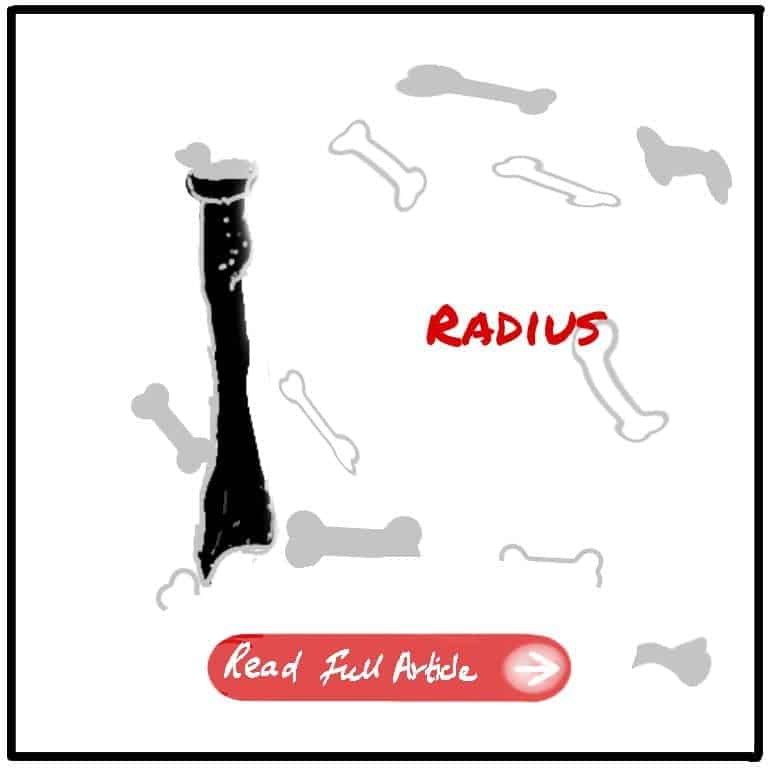
The radius bone, often overshadowed by its neighboring ulna, plays a crucial role in arm functionality and mobility. Located on the thumb side of the forearm, the radius bone is key to rotational movements and stability. In this article, we delve into the fascinating world of the radius bone, exploring its anatomy, function, and its significance in everyday activities. Join us as we unravel the hidden wonders of this essential bone and gain a deeper appreciation for its role in our arm’s remarkable capabilities.
Overview
The Radius is the lateral bone of the forearm. It is homologous with the tibia of the lower limb. The posterior interosseous nerve of the radial nerve wind around the neck of the radius. The brachial artery ends at the neck of the radius.
Side Determination
- 3 borders – anterior, posterior, interosseus
- 3 surfaces — anterior, posterior, lateral
- Anterior – anterior boarder
- Posterior – dorsal tubercle
- Medial – radial tuberosity
- Lateral – roughning for pronator teres attachment
- Superior – head of the radius
- Inferior – styloid process
Clinicals
01. Subluxatio of the head of radius — The Head of radius is pulled below. Common in children. The head is dislodged from the grip of annular ligament. This is also known as the nursemaid’s elbow.
02. Fracture proximal to pronator teres — The proximal fragment is supinated due to the action of biceps. The distal fragment is pronated.
03. Fracture distal to mid-shaft — The forearm remains in neutral position.
04. Fall on outstretched hand –
- In children — The distal radial epiphysis is displaced posteriorly
- In young adults — The shaft of the radius and ulna may fracture, along with scaphoid fracture.
- Elders —
- Colle’s fracture — Radius fracture 1 inch (2.54 cm) proximal to distal end, distal segment is displaced posteriorly.
- Smith’s fracture — The opposite of Colle’s fracture; the distal segment is displaced anteriorly.
05. Galeazzi’s fracture — Fracture of the distal part of the radius with dislocation of distal radio ulnar
joint and an intact ulna.
06. Fracture of the head of radius – Gives positive fat pad sign. When the head of the radius is fractured, the fluid fills the synovial cavity and elevate a small pad of fat within the coronoid and olecranon fossae and appear as areas of lucency in a lateral radiograph.


Leave a Reply