Muscles of the arm arrange in two compartments; anterior and posterior, which are
separated by intermuscular septa.
Anterior Compartment
The anterior compartment, also called the flexor compartment, is bordered anteriorly by the deep fascia, and posteriorly by the medial and lateral intermuscular septa and the Humerus in-between them. The blood supply to the entire anterior compartment is supplied by the axillary artery in the proximal end, and the brachial artery, which is the continuation of the brachial artery when it exits the axilla. Innervation to the anterior compartment is from the musculocutaneous nerve, which is a terminal branch of the lateral chord of the brachial plexus. This nerve is supplied by the nerve roots C5 to C7.
The specific attachments, innervation and blood supply of each muscle of the anterior compartment will be discussed in detail below.
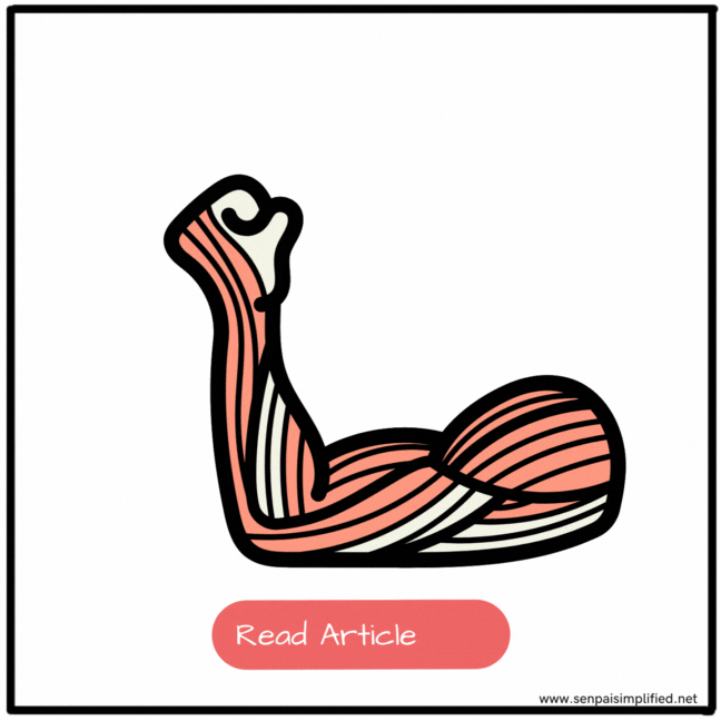
Biceps Brachii
This is the muscle everyone flexes to ‘show off their muscles’. The muscle has two heads. The long head and the short head. The long head arises from the supra glenoid tubercle of the scapula. The short head arises from the apex of the coracoid process of the scapula. The two heads then fuse together and insert into the radial tuberosity through a common tendon. Since this muscle is in the anterior compartment, it is innervated by the musculocutaneous nerve (C5,C6,C7).
The biceps brachii is a major flexor of the forearm, at the elbow joint. However, the primary function of the biceps brachii is supination of the forearm by rotating the radius laterally. Additionally, since both heads of the muscle pass through the shoulder joint, it is a weak flexor of the arm at the shoulder joint.
Coracobrachialis
The coracobrachialis originates from the apex of the coracoid process. This is an insertion that it shares with the long head of the biceps brachii muscle. They share this common tendon at insertion, and the tendon extends downwards as an intermuscular septum between the coracobrachialis and the biceps brachii. The coracobrachialis is inserted at a linear roughening on the medial side, in the mid-shaft of the humerus. This muscle is supplied by the nerve roots C5 and C6 of the Musculocutaneous nerve.
This is a flexor of the arm at the glenohumeral joint. It also facilitates a small degree of adduction. But coracobrachialis is not the primary muscle for either of these actions.
Brachialis
This is a flat muscle that gives girth to the arm. The brachialis originates from the anterior aspect of the humerus, and slightly from the medial and lateral aspects as well. It is also attached to the adjacent intermuscular septa at the posterior border of the anterior compartment. The muscle passes over the humerus and then inserts onto the tuberosity of the ulna. This muscle is innervated by the Musculocutaneous nerve (C5 and C6) and distally slight innervation from the radial nerve (C7) may be present.
This muscle is a powerful flexor of the forearm at the elbow joint. The main muscle that flexes the elbow is not the biceps brachii, it is this muscle. When the forearm is pronated, the Biceps are not acting. Hence, the brachialis muscle is the main muscle that flexes the pronated forearm along with some other muscles of the forearm.
Posterior Compartment
The posterior compartment is mainly occupied by the triceps brachii. . The posterior compartment is supplied by the profunda brachii artery. And it is innervated by the radial nerve (C5-T1)
There are some smaller muscles present distally near the elbow as well. Which are less important and probably out of your syllabus.
Triceps Brachii
This muscle almost entirely occupies the posterior compartment of the arm. As the name suggests, if biceps brachii has two heads, you guessed it… Triceps brachii has three heads. The long head originates from the infraglenoid tubercle of the scapula. The medial head originates from the medial side of the posterior surface of the Humerus. The lateral head arises from the lateral side of the posterior surface of the humerus. All three heads join together and fuse. So the joined muscle inserts into the olecranon process of the ulna. The Muscle is innervated by the Radial nerve (C6,C7,C8). Function
The main action of the triceps muscle is to extend the forearm at the elbow joint. However, since the long head is inserted in the scapula, the muscle has a small degree of extension and adduction at the glenohumeral joint as well.
Anconeus
This is a tiny muscle at the back of the elbow. It originates from the dorsal aspect of the lateral epicondyle of the humerus, just proximal to the common extensor origin of the forearm muscles. It inserts onto the lateral surface of the olecranon, and the posterior surface of the Ulna. It is innervated by a branch of the radial nerve called ‘nerve to Anconeus’ (C7,C8). Although insignificant, it functions as an extensor of the elbow.
Articularis cubiti
Some anatomists believe this to be a part of the tricep muscle itself, because it originates from the deep surface of the medial head of the triceps, distally. It inserts into the posterior surface of the capsule. Its function is to lift the capsule up when extending the elbow so that it doesn’t get pinched between the two bones. Isn’t it fascinating how we have evolved…
Clinical points
Popeye sign
Rupture of the biceps tendon gives the Popeye sign. Since the biceps muscle is a fusiform-shaped muscle, when the tending ruptures, it bulges up into a ball like shape, making it look like it is flexed when it is not. This sign has an interesting etymology. This is the classic sign you see in the cartoon character Popeye when he eats his spinach.


Leave a Reply