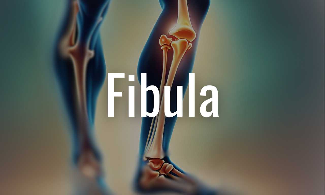
The fibula is one of the two bones in the lower leg, alongside the tibia. While the tibia bears most of the body’s weight, the fibula serves several important functions. Let’s explore the anatomy and clinical aspects of the fibula. Its upper limb counterpart ulna carries a lot of functional similarities to this.
Anatomy of the Fibula
We can describe the fibula by dividing it into the following components for ease. And, those compnents are, the head, neck, shaft, and lower end, also known as the lateral malleolus. So, let’s delve into each of these parts and look at their key features.
Head and Neck
- Origin for Muscles: The head of the fibula serves as an origin point for various muscles.
- Styloid Process: The styloid process, located in the head of the fibula, provides an attachment site for the biceps tendon.
- Common Peroneal Nerve: The common peroneal nerve transverses the neck of fibula. This is one of those cool features that you can touch and feel on your own leg. You can explore more of those in the palpable body structures article.
![]() Foot Drop:
Foot Drop:
Being so close to the skin means that it is far more vulnerable to injury. When this nerve gets damages, the whole anterolateral compartment of the leg loses innervation. Which results in a condition called foot drop.
Shaft
Subcutaneous Nature: The anterior and anteromedial surfaces of the fibula’s shaft are subcutaneous, meaning they lie just beneath the skin.
![]() Donor Site for Bone Grafts: Similar to the tibia, the fibula’s extensive subcutaneous surface makes it a suitable donor site for bone grafts in various surgical procedures.
Donor Site for Bone Grafts: Similar to the tibia, the fibula’s extensive subcutaneous surface makes it a suitable donor site for bone grafts in various surgical procedures.
Groove for Tendons: The posterior aspect of the lateral malleolus (lower end) features a groove that accommodates the tendons of the peroneus longus and peroneus brevis muscles.
Lower End (Lateral Malleolus)
- Articular Facet: The medial surface of the lateral malleolus articulates with the lateral surface of the talus bone, forming part of the ankle joint.
- Fibular Notch: The triangular area just above the articular facet fits into the fibular notch on the distal end of the tibia, where the two bones are joined by the distal end of the interosseous membrane.
- Malleolar Fossa: A pit or fossa on the posterior surface of the lateral malleolus serves as an attachment site for the posterior talofibular ligament associated with the ankle joint.
- Groove for Tendons: The posterior surface of the lateral malleolus contains a shallow groove that accommodates the tendons of the fibularis longus and fibularis brevis muscles.
![]() Non-Weight Bearing Bone:
Non-Weight Bearing Bone:
The fibula does not bear the majority of the body’s weight and is not directly involved in weight transmission. Its narrower shaft and non-weight-bearing function differentiate it from the tibia.
In conclusion, The fibula plays a significant role in muscle attachment, ankle joint function, and overall lower limb stability. Therefore, familiarity with its anatomy and clinical considerations assists healthcare professionals in providing effective care and treatment for patients.


Leave a Reply