In this article, we will delve into the cubital fossa or Chelidon, a triangular depression located in front of the elbow. It serves as a significant transitional area between the arm and the forearm, playing a crucial role in various clinical procedures. Let’s explore the cubital fossa in detail:
Introduction
The cubital fossa is a triangular depression that occupies the anterior aspect of the elbow. It is homologous to the popliteal fossa, which is found at the back of the knee joint. Understanding the boundaries and contents are essential for medical professionals working with the upper limb.
Boundaries of the Cubital Fossa
The cubital fossa is defined by the following boundaries:
- Lateral Boundary: Medial border of the brachioradialis muscle.
- Medial Boundary: Lateral border of the pronator teres muscle.
- Base: An imaginary line drawn between the medial and lateral epicondyles of the humerus.
- Apex: The meeting point of the medial and lateral boundaries.
- Floor: Composed of the supinator muscle (laterally) and the brachialis muscle (medially).
- Roof: From deep to superficial layers:
- Skin
- Superficial fascia
- Median cubital vein
- Lateral and median cutaneous nerves of the forearm
- Deep fascia
- Bicipital aponeurosis, separating the median cubital vein from the brachial artery and median nerve.
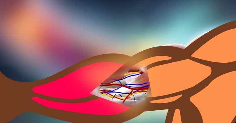
Contents of the Cubital Fossa
The cubital fossa contains important structures, arranged from lateral to medial. To remember the contents, you can use the mnemonic “R TAN” – Radial nerve, Tendon of biceps brachii, Brachial artery, Median nerve.
Nerves
The median nerve enters the cubital fossa between the heads of pronator teres and exits distally. It lies deep to the bicipital aponeurosis, coursing alongside the brachial artery. In the fossa, it gives off branches to surrounding muscles, contributing to forearm and hand movement. The radial nerve, on the other hand, traverses the cubital fossa’s roof, emerging from beneath the lateral border of the biceps brachii muscle. It supplies motor and sensory innervation to the posterior compartment of the forearm and hand. Additionally, the cubital fossa houses the lateral cutaneous nerve of the forearm, which emerges laterally from beneath the biceps tendon, providing sensory innervation to the anterolateral forearm. Note that the radial nerve is not a direct content of the cubital fossa but can be visualized when the brachioradialis muscle is retracted radially.
Biceps Brachii Tendon
The Biceps Brachii tendon inserts into the fossa. It attaches to the radial tuberosity. Near its insertion, the biceps tendon blends with the bicipital aponeurosis. This aponeurosis reinforces the roof of the cubital fossa.
Arteries
The brachial artery enters the fossa in the midline. It divides into radial and ulnar arteries halfway down the fossa. Transitioning to its forearm pathway, the brachial artery supplies the arm’s muscles. It courses alongside the median nerve within the cubital fossa. Deep to the artery lies the median nerve and brachialis muscle. The bicipital aponeurosis serves as a roof for the cubital fossa.
![]() Blood Pressure Measurements:
Blood Pressure Measurements:
During blood pressure measurements, healthcare providers commonly place the stethoscope over the brachial artery within the cubital fossa, to listen to the Korotkoff sounds.
![]() Palpation of the Brachial pulse:
Palpation of the Brachial pulse:
We can feel the brachial pulse by placing the finger on the cubital fossa. The brachial pulse is especially significant in neonatal examinations and other paediatric examinations, as the radial pulse may not be easily palpated due to the small size.
Veins
The cubital fossa hosts several veins, including the median cubital vein and its associated branches. Positioned superficially, these veins facilitate venous access for procedures like blood draws and intravenous therapy. The median cubital vein runs proximomedially from the cephalic to basilic veins. It lies superficial to the bicipital aponeurosis and communicates with deep veins. Dual venae comitantes usually accompany arteries, anastomosing freely with each other. The venous drainage of the upper limb is discussed in greater detail in a separate article.
![]() Phlebotomy/Venepuncture:
Phlebotomy/Venepuncture:
The median cubital vein is often preferred for venepuncture due to its accessibility and minimal risk of damage to surrounding structures. Healthcare professionals typically choose this site for blood draws or intravenous catheter insertions. The cubital fossa’s anatomical features, including the bicipital aponeurosis and surrounding musculature, provide support and stability during the procedure.
Lymph
Additionally, the supratrochlear lymph node lies in the superficial fascia over the upper part of the cubital fossa, above the trochlea. It receives lymph vessels from the third, fourth, and fifth fingers, the medial part of the hand, and the medial side of the forearm. The efferent lymph vessels travel to the axilla and enter the lateral axillary group of nodes.
To summarize the information about the cubital fossa, here’s a table:
| Aspect | Details |
|---|---|
| Boundaries | Lateral boundary: Medial border of the brachioradialis muscle |
| Medial boundary: Lateral border of the pronator teres muscle | |
| Base: Imaginary line between the medial and lateral epicondyles of the humerus | |
| Apex: Meeting point of the medial and lateral boundaries | |
| Floor | Supinator muscle (laterally) and brachialis muscle (medially) |
| Roof | From deep to superficial: Skin, superficial fascia, median cubital vein, lateral and median cutaneous nerves of the forearm, deep fascia, bicipital aponeurosis |
| Contents | Radial nerve (indirect content), Biceps brachii tendon, Brachial artery, Median nerve |


Leave a Reply