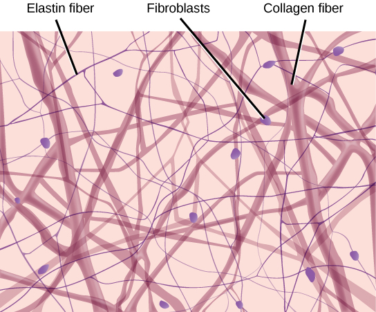
Connective tissue is the unsung hero of our body, holding everything together and ensuring smooth operation. It binds tissues and supports metabolic exchanges, thanks to its rich mix of collagen and glycosaminoglycans. This dynamic matrix not only maintains tissue strength but also defends against threats. Thus, by exploring its crucial role, we uncover its significance in various connective tissue disorders.
What is a Connective Tissue?
A tissue is A group or layer of cells that work together to carry out a specific role. A connective tissue connects tissues to other tissues. It also links tissues to structures and to the skeleton. It is a tissue which provides structural and metabolic support and functions as medium of exchange of nutrients and metabolites.
Functions of Connective Tissues
Connective tissue serves as biological packing material, filling the spaces between cells and other tissues. Loose connective tissue is found in the superficial fascia, the stroma of organs, and around nerves and blood vessels. In contrast, dense connective tissue is found in the dermis of the skin. It is also found in the capsules surrounding organs, tendons, and ligaments. Additionally, specialized types of connective tissue form rigid frameworks, like cartilage and bone. Connective tissue also has a metabolic role, storing fat in white adipose tissue and regulating body temperature in newborns through brown adipose tissue. Furthermore, it plays a crucial role in defending against pathogens and is essential for tissue repair.
Main components:
- Extracellular matrix
- Cells: Cells are responsible for the synthesis and maintenance of the EC matrix, storage, and metabolism of fats defence and immune functions. Most of the supporting tissues derive from embryonic Mesenchymal cells. These cells have an irregular spindle shape with slender processes. We discuss stem cells more broadly in a separate post.
- Ground Substance
Cells of supporting tissue
Supporting tissue includes three main types of cells, each with a specific role. Firstly, fibroblasts create the extracellular matrix. Meanwhile, tissue macrophages, mast cells, and leucocytes handle defense and immune functions. Lastly, adipocytes store and metabolize fats.


Fibroblasts
Fibroblasts are the most common cells in connective tissue. Their main job, thus, is to produce fibers and the ground substance of the extracellular matrix. Additionally, these cells are large and flattened, with oval-shaped nuclei and long cytoplasmic extensions. When active, the nucleolus becomes prominent, and fine chromatin granules appear. Well-developed ribosomes, rough endoplasmic reticulum, and a Golgi complex show their role in protein synthesis.
Defence cells of supporting tissue:
Macrophages

Macrophages come in two types: fixed (intrinsic) and wandering (extrinsic). Fixed macrophages include tissue macrophages and mast cells, while wandering ones are cells from the white blood series. These cells are ovoid with an oval nucleus that is indented on one side. The nucleus is irregular and contains heterochromatin.
In their active state, macrophages have abundant lysosomes, but these decrease in number when the cells are actively phagocytic. When active, they form pseudopodia, move in an amoeboid manner, and perform phagocytosis. Macrophages originate from blood monocytes, which migrate to peripheral tissues and become macrophages. They play a crucial role in the immune system..
Mast cells

Mast cells are oval or irregular in shape and are present in all types of supporting tissue. They are especially common beneath the skin, along the gastrointestinal and respiratory tracts, and around blood vessels. These cells resemble basophils, and their cytoplasm is full of large granules. When stained with toluidine blue, these granules turn red. The granules are membrane-bound and contain dense, amorphous material.
Mast cells play a key role in immune responses and pathological conditions, particularly in allergic reactions. However, they are absent in the spinal cord.
Leucocytes
Leucocytes in connective tissue look different from those in a blood smear. Neutrophils are rare, except in areas with acute or chronic inflammation. Eosinophils are found in large numbers within connective tissue. Lymphocytes are identified by their dense nucleus and a thin rim of cytoplasm.
Plasma cells

Plasma cells have a large amount of basophilic cytoplasm because of their rough endoplasmic reticulum and ribosomes. Inside the cytoplasm, there is a pale-stained area around the nucleus with a well-developed Golgi complex. These cells feature an eccentric nucleus with radiating chromatin that resembles a cartwheel. Plasma cells are active lymphocytes that produce antibodies and are larger than regular lymphocytes.
Adipocytes
Adipocytes are adapted for the storage of fat, and they are found in clusters in loose connective tissue. They are the main cell type in adipose tissue.

Extracellular matrix
The extracellular matrix gives supporting tissue its physical properties. It consists of organic material that forms the ground substance and various embedded fibers. Additionally, it holds structural glycoproteins that help cells interact with other components. Consequently, the nature of the extracellular matrix determines the differences among various types of supporting tissue.
Fibres of supporting tissue
Fibres are of three types:
- collagen (white) fibres
- elastic (yellow) fibres
- reticulin fibres
Collagen fibers
Collagen fibers exist in all types of supporting tissue. In fact, collagen is the most abundant protein in the human body. Additionally, these fibers are flexible and offer high tensile strength. Furthermore, fibroblasts secrete collagen into the extracellular matrix in the form of tropocollagen molecules..
Reticulin fibres
These fibres form the frame work in cellular organs such as liver, lymphoid tissue and bone marrow. The fine network of branching fibres anchor to the collagenous capsule and septae. These fibres are not clearly seen with hematoxylin and eosin. Reticulin fibres stain black with silver stains. In the process of formation of collagen, reticulin fibres are the earliest fibres to be produced.
![]() Ehlers-Danlos Syndrome (EDS)
Ehlers-Danlos Syndrome (EDS)
Ehlers-Danlos Syndrome (EDS) is a group of connective tissue disorders characterized by abnormalities in collagen fibers, which are crucial for maintaining the strength and elasticity of tissues. In EDS, defective collagen leads to a range of symptoms including hyperelastic skin, joint hypermobility, and a tendency to bruise easily. This condition highlights the critical role of collagen fibers in connective tissue, as they provide structural support and resilience.
Elastic fibres
Elastic fibres are thinner than collagen and exhibit no banding as in collagen. They are short branching fibres that form an irregular network.
Ground substance
It is an amorphous, transparent, hydrophilic material that exists as a semifluid gel, in which cells and fibers are arranged. Moreover, It consists mainly of glycosaminoglycans (mucopolysaccharides), primarily hyaluronic acid, linked to protein molecules to form proteoglycans, a type of mucoprotein. Additionally, fibrous proteins reinforce these mechanical proteins.
![]() Marfan Syndrome
Marfan Syndrome
Marfan syndrome stems from mutations in the gene coding for fibrillin-1, a key component of the extracellular matrix (ECM). This disruption in ECM integrity affects the ground substance, altering the structural framework of connective tissue. Consequently, individuals with Marfan syndrome exhibit weakened arterial walls, leading to aortic dilation and dissection, along with skeletal and ocular manifestations.
Tissue fluids, which are loosely associated with the ground substance, serve as a medium for exchanging nutrients, gases, and metabolites between cells and capillaries. Additionally, the ground substance forms a mechanical barrier to bacteria and plays an important role in preventing the spread of microorganisms. However, hyaluronidase present in some bacteria may aid in their spread. Moreover, the ground substance can act as a selective barrier to inorganic ions and charged molecules. Finally, structural glycoproteins consist of protein chains bound to branched polysaccharides.


Leave a Reply