When the bowel is obstructed in two adjacent places, it is known as a closed loop obstruction. In the case of a distal large bowel obstruction, if the ileocecal valve is competent, it creates a sealed loop.
The mechanics of closed loop obstruction
In any bowel obstruction, above the level of the obstruction, there is hyperperistaltism. This is when the rhythmic wave-like muscle contractions of the bowel become excessive and vigorous. Initially, in the first few hours, fermented gases from the microbiota of the colon would accumulate in the proximal bowel. This causes the distension of the loop. This causes an increase in pressure within the bowel. After 12 hours, there is fluid accumulation due to exaggerated intestinal secretion, which worsens the distension. This ultimately results in bowel perforation.
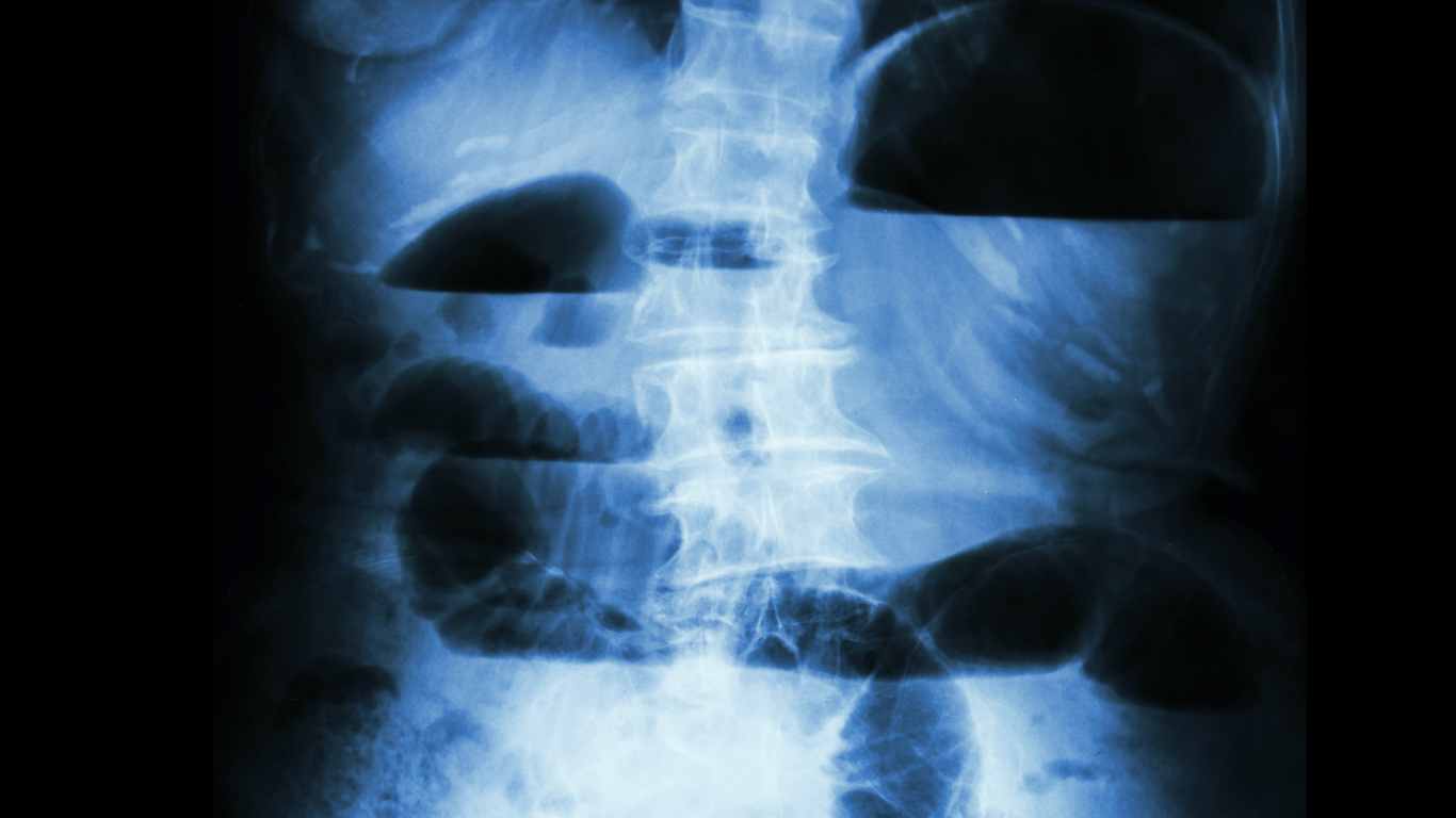
In a large colon closed loop obstruction with a competent ileocecal valve, the most vulnerable location for perforation is the caecum.
This can be explained using the physics concept Laplace’s Law, which states that the tension (T) in the wall of a hollow organ (like the caecum) is directly proportional to the pressure(P) inside and the radius(R) of the organ.
T=PR
To put it simply, with the pressure buildup due to the obstruction, the proximal bowel would distend. Since the caecum has the largest diameter in the colon, it requires the highest tension to withstand distension. Therefore, it is most likely to distend and potentially perforate.

Let’s apply this to a real life clinical scenario:
Imagine you have a patient with abdominal pain, distension, and absolute constipation. (Absolute constipation is when the patient is passing no flatus or faeces.) These are cardinal signs of bowel obstruction. Vomiting is also a cardinal sign. Small bowel obstructions more prominently cause vomiting. While patients with large bowel obstructions can experience vomiting, this rarely occurs and indicates a late stage.
The urgency in which you have to treat this patient depends on their clinical status. If there was an incompetent ileocoecal valve seen in 20-30% of cases, the colon can decompress into the small bowel. The need for intervention is not as urgent as in the case of a closed-loop obstruction.
If this patient develops localised right iliac fossa pain, that is a major red flag.
As mentioned above, the caecum, due to its diameter, is more likely to perforate due to increased pressure inside the bowel lumen. This impending perforation manifests as right illiac fossa pain, and clinicians should act quickly.
In the colon, the other cause of closed loop obstruction is in the case of volvulus. It is when the colon twists on itself. This most commonly occurs in the sigmoid colon due to its naturally long and mobile mesentery. Here, the main feared complication is ischaemia of the bowel due to vascular obstruction. The vascular pedicle, which carries the arteries of that specific segment of bowel, is compressed.
How is it managed clinically?
First and foremost, the patient needs to be resuscitated with fluids and electrolyte repletion, especially potassium.
We keep the patient nil by mouth to rest the gut. We insert a NG tube to decompress distended bowel and prevent aspiration in case of vomiting. Go for radiological imaging (CECT is preferred) if the patient is stable.
If the obstruction is caused by stricture/ tumour midline laparotomy is indicated and subtotal colectomy maybe needed.
Decompression of the sigmoid volvulus can be done using Flexible Sigmoidoscopy or colonoscopy. This also allows us to visualise any ischemic areas in the bowel wall, which is a significant advantage. It is both diagnostic and therapeutic. When the point of rotation is reached, there is dramatic release of the obstruction. The surgeon relieves the blockage and then leaves a tube in place for 48 hours. The team performs emergency surgery only if they find signs of tissue ischemia.
References
1. https://www.ncbi.nlm.nih.gov/books/NBK441925/
2. Mbengue, A., Ndiaye, A., Soko, T.O., Sahnoun, M., Fall, A., Diouf, C.T., Régent, D. and Diakhaté, I.C., 2015. Closed loop obstruction: pictorial essay. Diagnostic and Interventional Imaging, 96(2), pp.213-220.


Leave a Reply