The brachial plexus is a complex network of nerves that originates from the anterior (ventral) primary rami of C5, C6, C7, C8, and T1 spinal nerves. It plays a vital role in the innervation of the upper limb. Let’s explore the anatomy and components of the brachial plexus in more detail:
Anatomy of the Brachial Plexus
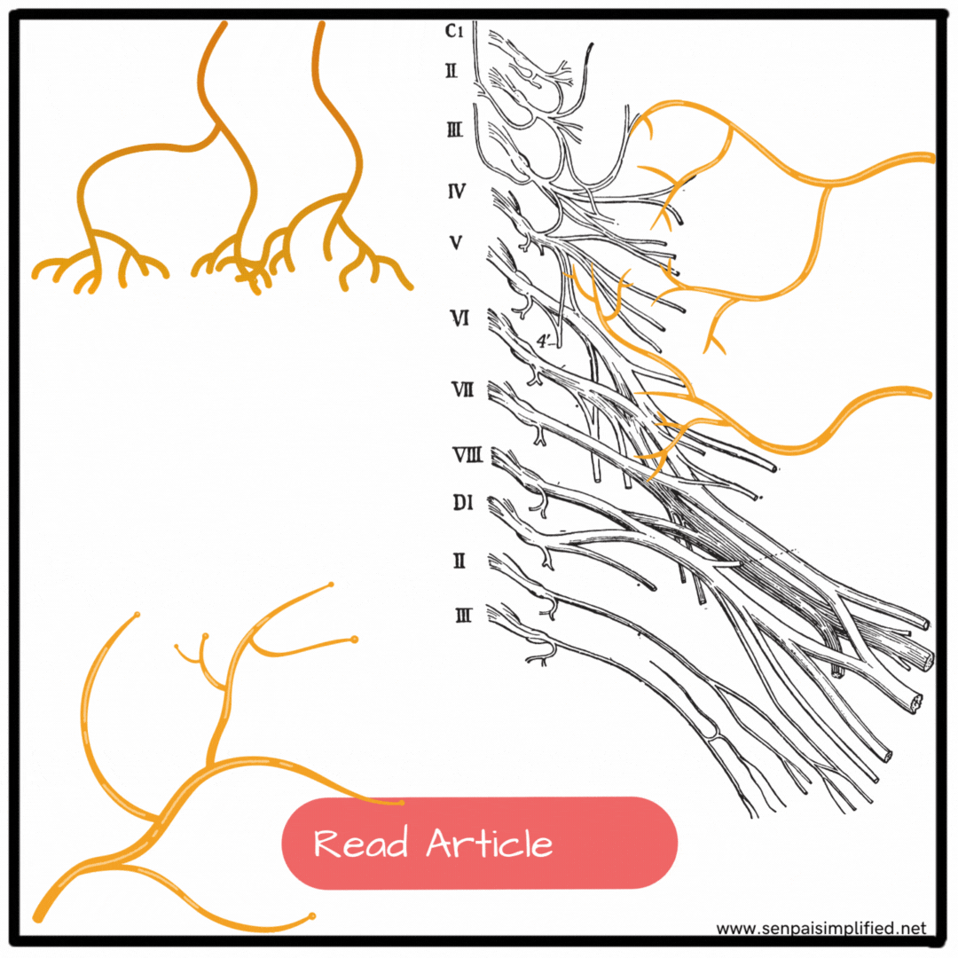
The brachial plexus is formed by the union of the anterior rami of the 5th, 6th, 7th, and 8th cervical and the 1st thoracic spinal nerves. It is located in the posterior triangle of the neck and can be divided into roots, trunks, divisions, and cords.
– Roots: The roots of C5 and C6 unite to form the upper trunk, C7 continues as the middle trunk, and C8 and T1 unite to form the lower trunk.
– Trunks: The trunks traverse the posterior triangle of the neck.
– Divisions: The divisions lie behind the clavicle.
– Cords: The cords of the brachial plexus are formed by the union of specific divisions. The lateral cord is formed by the union of the anterior divisions of the upper and middle trunks, the medial cord continues as the anterior division of the lower trunk, and the posterior cord is formed by the union of the posterior divisions of all three trunks.
These cords mix with each other to form Branches.
Mnemonic : The order of these different components of the brachial plexus is remembered by the mnemonic:
Randy Travis Drinks Cold Beer
Randy – Roots
Travis – Trunks
Drinks – Divisions
Cold – Cold
Beer/ Beverages – Branches
Branches of the Brachial Plexus
The brachial plexus gives rise to various branches that supply motor and sensory innervation to different structures in the upper limb. Let’s explore some of the key branches:
Branches arising from the Roots
- – Dorsal scapular nerve (C5)
- – Long thoracic nerve (C5, C6, and C7)
Upper Trunk
– Nerve to subclavius (C5 and C6)
– Suprascapular nerve (supplies the supraspinatus and infraspinatus muscles)
Lateral Cord:
– Lateral pectoral nerve
– Musculocutaneous nerve
– Lateral root of the median nerve
Medial Cord
– Medial pectoral nerve
– Medial cutaneous nerve of arm and medial cutaneous nerve of forearm
– Ulnar nerve
– Medial root of the median nerve
Posterior Cord:
– Upper and lower subscapular nerves
– Thoracodorsal nerve
– Axillary nerve
– Radial nerve
Branches in the Axilla
Within the axilla, several branches of the brachial plexus emerge to innervate specific structures:
– The nerve to the subclavius (C5 and C6) supplies the subclavius muscle.
– The long thoracic nerve (C5, C6, and C7) descends over the lateral surface of the serratus anterior muscle, which it supplies.
– The lateral pectoral nerve supplies the pectoralis major muscle.
– The musculocutaneous nerve supplies the coracobrachialis muscle and pierces it.
– The lateral root of the median nerve is a continuation of the lateral
Brachial Plexus Branches
Here is a comparison table summarizing the branches of the brachial plexus and their functions:
| Branches | Root | Distribution |
|---|---|---|
| Roots: | ||
| Dorsal scapular nerve | (C5) | Rhomboid minor, rhomboid major, levator scapulae muscles |
| Long thoracic nerve | (C5, 6, 7) | Serratus anterior muscle |
| Upper Trunk: | ||
| Suprascapular nerve | (C5, 6) | Supraspinatus and infraspinatus muscles |
| Nerve to subclavius | (C5, 6) | Subclavius |
| Lateral Cord: | ||
| Lateral pectoral nerve | (C5, 6, 7) | Pectoralis major muscle |
| Musculocutaneous nerve | (C5, 6, 7) | Coracobrachialis, biceps brachii, brachialis muscles; supplies skin along lateral border of forearm |
| Lateral root of median nerve | (C5, 6, 7) | See medial root of median nerve |
| Posterior Cord: | ||
| Upper subscapular nerve | (C5, 6) | Subscapularis muscle |
| Thoracodorsal nerve | (C6, 7, 8) | Latissimus dorsi muscle |
| Lower subscapular nerve | (C5, 6) | Subscapularis and teres major muscles |
| Axillary nerve | (C5, 6) | Deltoid and teres minor muscles; upper lateral cutaneous nerve of arm supplies skin over lower half of deltoid muscle |
| Radial nerve | (C5, 6, 7, 8; T1) | Triceps, anconeus, part of brachialis, extensor muscles of forearm, skin on dorsum of hand and fingers, and articular branches to elbow, wrist, and hand |
| Medial Cord: | ||
| Medial pectoral nerve | (C8; T1) | Pectoralis major and minor muscles |
| Medial cutaneous nerve of arm | (C8; T1, 2) | Skin of medial side of arm |
| Medial cutaneous nerve of forearm | (C8; T1) | Skin of medial side of forearm |
| Ulnar nerve | (C8; T1) | Flexor carpi ulnaris and medial half of flexor digitorum profundus, intrinsic hand muscles, and skin of medial hand and fingers |
| Medial root of median nerve (with lateral root) forms median nerve | (C5, 6, 7, 8; T1) | Pronator teres, flexor carpi radialis, palmaris longus, flexor digitorum superficialis, and more |
This table provides an overview of the main branches of the brachial plexus, their associated functions, and the muscles or areas they innervate.
Understanding the branches of the brachial plexus and their functions is essential in diagnosing and treating conditions affecting the upper limb. It enables healthcare professionals to accurately assess nerve-related
Clinical Considerations
The brachial plexus is susceptible to various injuries, which can lead to specific clinical conditions. Let’s explore two common brachial plexus injuries and their clinical features:
01. Erb’s Paralysis:
- Site of Injury: The region of the upper trunk of the brachial plexus, known as Erb’s point.
- Cause of Injury: Erb’s paralysis often occurs due to undue separation of the hand from the shoulder, which can happen during birth injuries, falls on the shoulder, or even during anesthesia.
- Nerve Roots Involved: It primarily affects the C5 nerve root, with partial involvement of the C6 nerve root.
- Muscles Paralysed: Erb’s paralysis results in the paralysis of specific muscles, including the biceps, brachialis, brachioradialis, deltoid, supraspinatus, infraspinatus, and supinator muscles.
- Deformity: The characteristic deformity associated with Erb’s paralysis is known as Policeman’s tip hand or Porter’s tip hand. In this condition, the affected arm hangs by the side, adducted, and medially rotated. The forearm remains extended and pronated.
- Disability: Erb’s paralysis leads to the loss of abduction and lateral rotation of the arm, as well as the inability to flex and supinate the forearm. Jerks of the biceps and supinator muscles are lost, and there is a loss of sensation over a small area on the lower half of the deltoid.
02. Klumpke’s Paralysis:
- Site of Injury: Klumpke’s paralysis affects the lower trunk of the brachial plexus.
- Cause of Injury: It is commonly caused by undue abduction of the arm, such as holding a support while falling from above, or as a result of birth injuries.
- Nerve Roots Involved: The main nerves involved in Klumpke’s paralysis are T1 (majority) and partially C8.
- Muscles Paralysed: This condition results in the paralysis of intrinsic muscles of the hand (controlled by the T1 nerve root) and ulnar flexors of the wrist and fingers (controlled by the C8 nerve root).
- Deformity: Klumpke’s paralysis leads to a characteristic deformity known as claw hand. It occurs due to the unopposed action of the long flexor and extensor muscles of the fingers.
- Disability: The primary disability associated with Klumpke’s paralysis is a claw hand, where the fingers are hyperextended at the metacarpophalangeal joints and flexed at the interphalangeal joints. Additionally, there may be cutaneous anaesthesia and analgesia over a narrow zone along the ulnar border of the hand and forearm.


Leave a Reply