Similar to the arm, the forearm is also divided into anterior and posterior muscle compartments. In this article, we will look at the attachments, innervation and action of all of those muscles in the anterior compartment of the forearm.
Superficial Layer
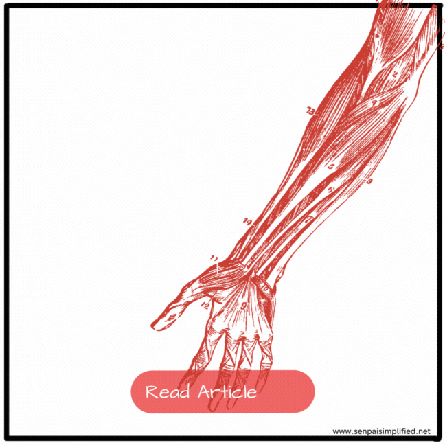
There are a lot of muscles in the hand. But muscles in each compartment and layer shares innervation blood supply and sometimes even origins and insertions with the adjacent muscles, with few exceptions. Similarly, all the muscles in all three layers (superficial, intermediate and deep) of the anterior compartment of the arm are given blood supply by the radial and ulnar arteries, which travel in the anterior compartment. All the muscles in the superficial layer of the anterior compartment of the arm, are innervated by the median nerve, except for the Flexor carpi ulnaris, which is innervated by the ulnar nerve. The origin, insertion and function of each muscle will be discussed separately.
Flexor Carpi Ulnaris
The flexor carpi ulnaris has two heads. The humeral head, originates from the medial epicondyle of the humerus. The ulnar head arises from the olecranon and the posterior border of the Ulna. It gets inserted into the pisiform bone and then, via pisohamate and pisometacarpal ligaments, attach to the hamate and the base of the 5th metacarpal respectively.
The pisiform bone is innervated by the ulnar nerve (C7,C8,T1). This is an exception from the rest of the muscles of the superficial layer of the anterior compartment of the forearm, because the rest of the muscles are innervated by the median nerve.
The flexor carpi ulnaris flexes and adducts the wrist joint.
Palmaris Longus
This muscle is only present in some individuals. The presence of the palmar is longus can be elicited by bringing the tips of the first and fifth digits together. The string like muscle will bulge out at the wrist.

The Palmaris longus arises from the medial epicondyle of the humerus, at the common flexor origin. The muscle gets inserted to the palmar aponeurosis of the hand.
Since this muscle is in the superficial layer of the anterior compartment, It is innervated by the Median nerve. Specifically, the C7 and C8 nerve roots. The exact nerve roots supplying each muscle will not be expected to be memorized in most medical schools. But they are mentioned here only for completion’s sake.
The Palmaris longus flexes the wrist joint. Since the palmar aponeurosis, which this muscle is attached to, is anchored to the skin of the hand, the contraction of the muscle resists shearing forces when gripping.
Flexor Carpi Radialis
The flexor carpi radialis originates from the medial epicondyle of the humerus, from the common flexor origin. Then it gets attached inferiorly at the bases of the second and third metacarpal bones. This muscle is also innervated by the median nerve.
The flexor carpi radialis flexes and abducts the hand at the wrist joint.
Pronator Teres
Similar to the flexor carpi ulnaris, the pronator teres also has two heads. The humeral head arises from the medial epicondyle and the adjacent supra-epicondylar ridge. The ulnar head arises from the medial side of the coronoid process. Now look again where the ulnar head of the flexor carpi ulnaris originates from, and compare. The muscle traverses diagonally and inserts onto a roughening on the lateral surface of the midshaft of the radius. This is the principle pronator of the forearm. However, this muscle is at its mechanical advantage, when the elbow is partially flexed because this muscle passes through the elbow joint as well.
Intermediate Layer
Flexor Digitorum Superficialis
The intermediate layer only consists of a single muscle, the flexor digitorum superficialis (FDS). You might think because this muscle is named ‘superficialis’, it must be in the superficial layer, but this muscle is underneath those muscles separating them from the deep muscles.
The flexor digitorum superficialis also has two heads: The Humero-ulnar head, which arises from the medial epicondyle of the humerus. And the adjacent margin of the coronoid process. The radial head arises from the oblique line of the radius. Distally the two heads come together, and divide again into 4 tendons that attach to the palmar surfaces of the middle phalanges of the digits 2 -5 (index, middle, ring and little fingers).
The flexor digitorum superficialis flexes the proximal interphalangeal (PIP) joints of the digits 2 through 5. It can also flex the metacarpo-phalangeal joints of those fingers.
Deep Layer
Since this is in the anterior compartment, blood is supplied by the Radial and ulnar arteries. All the muscles are basically innervated by the median nerve Except for FDP which has a mixed innervation.
Flexor Digitorum Profundus
This is the counterpart of the flexor digitorum superficialis. Some say the word profundus is related to the word profound, which is a misnomer. The actual Latin word profundus means deeps [Pro (Before) + Fundus (Bottom)]. Where there’s a superficialis, there is always a profundus.
The flexor digitorum profundus (FDP) originates from the anterior and medial surfaces of the ulna, and the medial surfaces of the interosseous membrane. The muscle divides into four tendons similar to the FDS. A tendon attaches to palmar surfaces of the distal phalanges 2 through 5. The medial half of the muscle is innervated by the median nerve. Conversely, the lateral half is innervated by the ulnar nerve (C8,T1).
The function of the FDP is, it flexes the distal interphalangeal (DIP) joints of the index, middle, ring and little fingers. It can also flex the metacarpo-phalangeal joints, proximal interphalangeal joints, and the wrist joint.
Flexor Pollicis Longus
The flexor pollicis longus is the longer counterpart of the flexor pollicis brevis, which is a muscle of the hand. Where there is a longus, there must always be a brevis, and vice versa.
This muscle originates from the anterior surface of the radius and the radial half of the interosseous membrane, and then gets inserted distally, to the palmar surface of the base of the distal phalanx of the thumb. This muscle is innervated by the anterior interosseous nerve (C7, C8), which is a branch of the median nerve.
The flexor digitorum longus muscle flexes the interphalangeal joint of the first digit (thumb). Remember that the thumb has only two phalanges and, hence, only one interphalangeal joint. It is a common rookie mistake to refer to this interphalangeal joint as the DIP or PIP joint, This can also flex the metacarpophalangeal as it passes through this as well.
Pronator Quadratus
The pronator quadratus is a quadrangle shaped muscle. It originates from the linear edge on the distal surface of the ulna, and the insertion is on the distal anterior surface of the radius. It is innervated by the anterior interosseous nerve (C7, C8). This is a pronator of the radius, specially when the elbow is extended, this is the main pronator of the forearm.


Leave a Reply