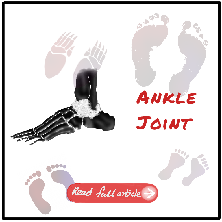
In this article we will discuss the type, articulations stabilizing factors and movements of the ankle joint. Some clinical points relating to the ankle joint will also be discussed.
Joint Type
This is a Hinge type joint with movement along only one axis. Do not confuse this with the subtalar and talo-calcaneo-navicular joint.
Articular surface
The articular Surfaces that form this joint are,
- Lower end of the tibia, including the medial malleolus
- The lateral malleolus of the fibula
- The inferior Transverse Tibio-fibular ligament
These together form a sort of deep socket That forms the upper articular surface. The lower articular surface is formed by articular areas on the upper medial and lateral aspects of the talus.
Stabilizing Factors
The stability of The joint is ensured by close interlocking of articular surfaces, strong collateral ligaments, and the tendons that cross the joint.
Tendons
There are 4 tendons in front, –
- Tibialis Anterior
- Extensor Hallucis Longus
- Extensor Hallucis Brevis
- Peroneus Tertius
3 Tendons in the Posteromedial Aspect and
- Tibialis Posterior
- Flexor Digitorum Longus
- Flexor Hallucis Longus
2 on the posterolateral aspect.
- Peroneus Longus
- Peroneus Brevis
Articular Surface
Another Factor that increases the stability of the joint is its depth and concavity of the upper articular surface. The first and more apparent factor is the downward projection of medial and lateral malleoli.
The other is the inferior transverse Tibio-fibular ligament that bridges between the Tibia and fibula behind the talus. The Forward pull of the tendons and the weight of the foot when the heel is raised tend to displace the tibia and fibula anteriorly over the talus. This is prevented by The wedge shape of the Talus. It is wider anteriorly, so that the malleoli don’t slip forward.
The posterior border of the lower end of the tibia is prolonged downwards. The presence of the inferior transverse Tibiofibular ligament. That we discussed earlier also helps in the forward displacement of tibia and fibula over the talus. And also parts of the Medial and lateral collateral ligaments help in this stability
Ligaments
The Fibrous Capsule
It surrounds the joint Entirely. But it’s relatively weak anteriorly and posteriorly. Since it is a synovial joint, the capsule is lined by synovial membrane,
It’s attached all around the articular margins, with 2 exceptions,
- Posteriorly, it is attached to the inferior transverse Tibiofibular ligament.
- And antero-inferiorly it is attached to the Dorsum of the neck of talus at some distance away from the trochlear surface.
The loose and thinness of the capsule anteriorly and posteriorly allows for hinge movements.
Collateral ligaments.
Unlike the anterior and posterior aspects, the lateral and medial sides have to be highly enforced and immobile to restrict the joint to hinge movement only.
The medial Ligament or the Deltoid ligament.
This is a Triangular-shaped ligament and hence given the name ‘Deltoid’ ligament.
It’s composed of 3 parts
- Anterior or tibio navicular Fibres. It attaches to the tuberosity of the navicular bone. And to the medial margin of the spring ligament.
- The middle, or Tibiocalcanean Fibres, are attached to the entire length of the sustenaculum tali.
- The posterior, or posterior tibiotalar Fibres, is attached to the medial tubercle of the posterior process of the talus.
The deltoid ligament is crossed by tendons of tibialis posterior and flexor digitorum longus.
The Lateral ligament
This ligament consists of three brands as follows.
- Anterior talofibular ligament
- Posterior talofibular ligament
- Calcaneofibular ligament
Movements
The movements allowed by the ankle joint are dorsiflexion and plantar flexion. This is not to be confused with the Subtalar and talo-calcaneo-navicular joints that allow inversion and eversion of the foot.
Innervation and Blood Supply
According to the Hilton’s law, This joint is also supplied by the nerves that pass through it, which are deep peroneal and tibial nerves. And Blood supply is by anterior tibial posterior tibia and talotibial arteries.
Clinical Points
Dislocations of the ankle joint are very rare because the ankle joint is very stable Due to all the factors I mentioned previously in the video. Acute sprains of the medial ankle may occur due to excessive eversion, but abduction sprains of the ankle are almost always sprains of the subtalar joint. The true sprains of the medial ankle joint may occur with the tear of the strong deltoid ligament
The lateral collateral ligament is far more frequently affected in ankle sprains because the medial collateral ligament is stronger, and also because you are more likely to forcibly invert your ankle rather than evert. An abduction external-rotation injury of the ankle joint may cause 1st 2nd or 3rd degree Potts fracture


Leave a Reply