The heart has a unique electrical system that allows it to pump even when the brain is dead. This fascinating effect is enabled by several physiological phenomena, which are discussed in this article.
Resting membrane potential of cardiac muscles
The resting membrane potential (RMP) of the cardiac muscle is different to that of neurons and skeletal muscles. The cardiac muscle RMP is around -90mV. It is caused by K+ leak channels and Na+/K+/ATPase pump. The cardiac resting membrane potential is dependent on the extracellular concentration of K+, therefore, variations of the Potassium ion concentration in plasma affect the RMP.
In hyperkalaemia, the inside of the cell becomes more negative compared to the outside. Hence, the cell become more difficult to get depolarized. In hyperkalaemia, the inside of the cell becomes less negative. Hence, inside the cell becomes less negative which make it easier to get depolarized. The depolarization of the resting membrane potential beyond the threshold gives rise to an action potential (AP)
Action potential of ventricular muscles
The cardiac muscle action potential has 5 phases :
- Phase 0 (Rapid depolarization)
- Phase 1 (Partial repolarization / initial rapid repolarization)
- Phase 2 (Plateau phase)
- Phase 3 (Final slow repolarization)
- Phase 4 (RMP)
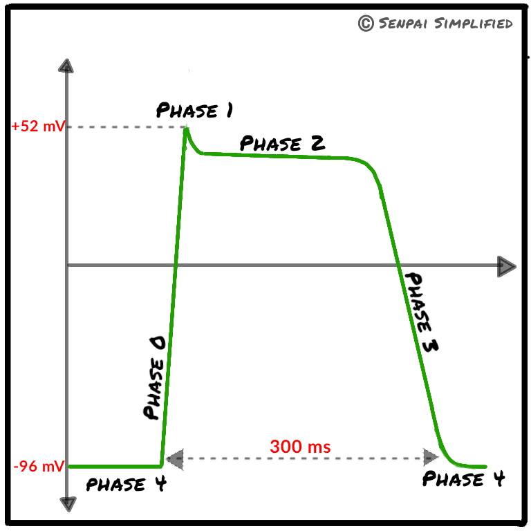
The cardiac action potential lasts about 300 milliseconds, which is significantly longer than the durations of skeletal muscles and nerve cells (2-5 ms and 1ms respectively). The magnitude of the cardiac action potential is determined by the concentration of Na+ in the Extracellular fluid(ECF).
Let’s take a look at the events taking place in each phase of the cardiac action potential.
Phase 0
Voltage gated Na+ channels open and influx of Na+ occurs. The membrane potential depolarizes to about +20mV. This phase lasts for about 2ms.
Phase 1
Transient K+ channels open and voltage gated Na+ channels close. This halts the influx of Na+ into the cell and starts sending K+ ions out of the cell. This causes a partial repolarization.
Phase 2
Voltage gated L- type Ca2+ channels open and delayed rectifier K+ channels also open. The resulting Ca2+ influx causes the membrane potential to get depolarized, meanwhile the K+ efflux causes the membrane potential to get repolarized. The tug of war between these phenomena keeps the membrane potential constant in a plateau phase.
Phase 3
Various K+ channels are kept open, but voltage gated Ca2+ channels close. The influx of Ca2+ stops, but the efflux of K+ continues, resulting in the final membrane repolarization.
Phase 4
The repolarized membrane potential reaches the RMV of -90mV.
Excitation – contraction coupling
AP of cardiac muscles is coupled with contraction. AP of cardiac muscle is an electrical response and the contraction of cardiac muscle is a mechanical response. Contraction begins just after the start of depolarization. Lasts 1.5 times as long as the action potential. The mechanism of contraction is described in the following steps
- AP spreads inside the muscle cell via T- tubules
- Causes entry of Ca2+ from ECF to ICF. (Via DHP channels)
- Release Ca2+ from SR.
- Ca2+ binds with troponin c
- Take away tropomyosin filaments from myosin binding sites on actin.
- Myosin binds with actin.
- Initiate contraction.
Releasing of Ca2+ from the sarcoplasmic reticulum (SR) is triggered by the entry of ECF Ca2+. Cardiac muscle needs influx of Ca2+ from ECF because the stored Ca2+ in SR is insufficient. T- tubules wider in cardiac muscle than in skeletal muscle because of this. When ECF Ca2+ concentration decreases, the cardiac muscle contraction is reduced.
The mechanism of relaxation of the cardiac muscle occurs by efflux of Ca2+. Ca2+ pumps out from troponin C (To SR / To ECF). This is an energy requiring process. The pump to SR Use energy from Ca2+– ATPase pump. The pump to ECF – Uses Na+ – Ca2+ exchanger and Na+– K+ ATPase pump.


Leave a Reply