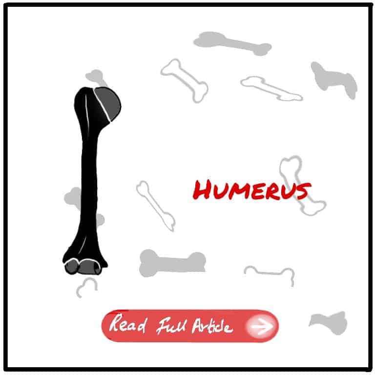
The humerus, the long bone in the upper arm, forms a critical link between the shoulder and the elbow. As the largest bone in the upper body, the humerus provides structural support and enables a wide range of arm movements. In this article, we delve into the fascinating world of the humerus, exploring its anatomy, function, and its crucial role in facilitating everyday activities. Join us as we uncover the remarkable features and significance of this essential bone.


Overview
Humerus is the longest bone of the upper limb. It articulates with the glenoid cavity of the scapula at the shoulder joint and with the radius and ulna at the elbow. It consists of a head, neck shaft and the distal end has a capitulum and trochlea. The head is 1/3rd of a sphere which is facing medially, upward and backward. It is separated from the shaft by surgical neck.
Greater and lesser tubercles are separated from the head by the anatomical neck and
each tubercle is separated from each other by intertubercular sulcus (bicipial groove). The shaft is circular in above and triangular in below. The lower end of the humerus is angled forward 40° from the shaft. The axillary nerve and circumflex humeral vessels wind around the surgical neck. Radial nerve with profunda brachii vessels at the radial groove of the shaft. The ulnar nerve passes behind then below the medial epicondyle.
Side Determination
- Anterior — lesser tubercle, coronoid fossa, radial fossa, capitulum(only present in the anterior side whereas the trochlea present in both anterior and posterior sides)
- Anteromedial — nutrient foreman
- Anterolateral — deltoid tuberosity.
- Posterior — Olecranon fossa, radial groove
- Medial — head of the humerus
- Lateral — greater tubercle
- Superior — head, tubercles
- Inferior — trochlea, capitulum, epicondyles, supracondylar ridges
Clinicals
Fracture of the surgical neck – Prone damage to axillary nerve since it winds around the surgical neck. Damage to the axillary nerve causes loss of sensation on the regimental badge area on the lower lateral surface of the deltoid.
Mid-shaft fracture — Prone damage to radial nerve and profunda brachii vessels.
Lower end shaft fracture — prone damage to ulna nerve
Supracondylar fracture — Prone damage to brachial artery and median nerve. Elbow become unduly prominent. Doesn’t interrupt the equilateral triangular relationship between the olecranon process, and the medial and lateral epicondyles. Decrease of the angulation which indicate backward displacement of the
distal end.
Volkmann’s ischemic contracture — lower end of the proximal fragment damages the
brachial artery results in ischemia of the forearm muscles, which leads to fibrosis and
contraction of long flexors and extensors.
Wrist — flexed (As flexors are bulkier than extensors)
Metacarpo-phalangeal joint — extended (As insertion of extensor tendons into metacarpo-phalangeal joints)
Interphalangeal joints-flexed (As insertion of long flexor tendons into IPJs)


Leave a Reply