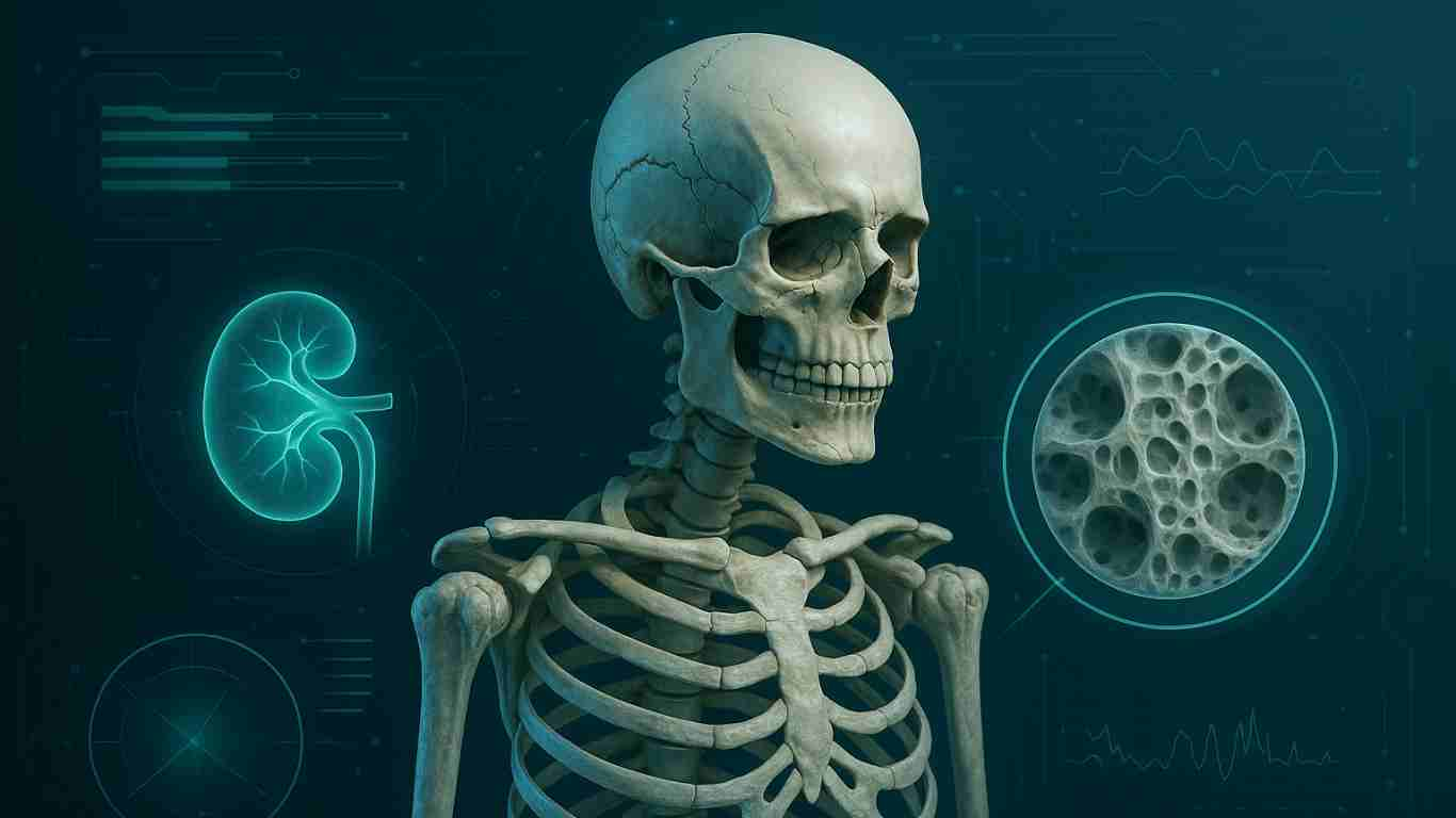
Chronic Kidney Disease-Mineral and Bone Disorder (CKD-MBD) is a complex and systemic complication that arises as kidney function declines. [1] It’s a web of interconnected abnormalities. It involves mineral metabolism (calcium, phosphorus), hormones (parathyroid hormone, vitamin D, and Fibroblast Growth Factor 23), and bone health. But, these abnormalities are not a single disease. They lead to significant cardiovascular consequences. [2] The mechanism is a cascade of events. In other words, it is a vicious cycle. Ultimately, the body’s attempts to compensate for failing kidneys cause more harm. [3]
The root of the problem lies in the kidneys’ diminished ability to do their vital functions. As the glomerular filtration rate (GFR) falls, the body’s intricate system for maintaining mineral and bone homeostasis begins to unravel.
The Cascade of Dysregulation:
The pathogenesis of CKD-MBD can be understood as a sequence of interconnected events:
Phosphorus Retention and the Rise of FGF-23:
In the early stages of CKD, the kidneys’ ability to excrete phosphate becomes impaired. As a result, this leads to a tendency towards phosphate retention in the blood. When phosphate levels increase even slightly, bone cells called osteocytes release a hormone. This hormone is called Fibroblast Growth Factor 23 (FGF-23). [4] FGF-23 acts as an early compensatory mechanism by signaling the remaining functional nephrons to increase phosphate excretion. [5]
The Double-Edged Sword of FGF-23 and Vitamin D Deficiency:
While initially helpful, rising FGF-23 levels have a significant downside. FGF-23 also inhibits the enzyme 1-alpha-hydroxylase in the kidneys. [6] This enzyme is crucial for converting inactive vitamin D (25-hydroxyvitamin D) into its active form, calcitriol (1,25-dihydroxyvitamin D). [6] So, as FGF-23 levels surge, the production of active vitamin D plummets. [6] This decline in calcitriol has two major consequences:
- Reduced Intestinal Calcium Absorption: Calcitriol is the primary hormone responsible for facilitating the absorption of calcium from the gut. [1] With less calcitriol, intestinal calcium absorption decreases, leading to lower levels of calcium in the blood (hypocalcemia). [1]
- Loss of a Key Regulator: Calcitriol normally acts as a negative regulator of parathyroid hormone (PTH) secretion. [7]
The Onset of Secondary Hyperparathyroidism:
Low blood calcium levels and decreased active vitamin D stimulate the parathyroid glands. This stimulation leads to an increase in the production and secretion of Parathyroid Hormone (PTH). This condition is known as secondary hyperparathyroidism.
The Destructive Actions of Excess PTH:
PTH’s primary role is to raise blood calcium levels. In a desperate attempt to normalize calcium, the persistently high levels of PTH in CKD lead to:
- Bone Resorption: PTH stimulates osteoclasts, the cells responsible for breaking down bone tissue. [7] This process, known as bone resorption, releases calcium and phosphorus from the skeletal reserves into the bloodstream. As a result, this constant breakdown weakens the bones. It leads to a condition called renal osteodystrophy. This condition is characterized by increased fracture risk and bone pain. [1]
- Increased Renal Calcium Reabsorption: PTH also acts on the kidneys. It increases the reabsorption of calcium. This further attempts to boost blood calcium levels. [7] The Vicious Cycle Intensifies: The increased bone resorption is driven by high PTH. It releases more phosphorus into the blood. This further exacerbates the initial problem of phosphate retention. This stimulates more FGF-23 production. This leads to an even greater suppression of active vitamin D. It also causes a more potent stimulus for PTH secretion. This creates a relentless and damaging feedback loop.
The Consequences: A Triad of Trouble
This complex interplay of hormonal and mineral imbalances culminates in the clinical manifestations of CKD-MBD, which can be broadly categorized into a triad of problems:
- Abnormalities of Mineral Metabolism: This includes high levels of phosphorus (hyperphosphatemia). It also involves low or sometimes high levels of calcium (hypocalcemia or hypercalcemia). Additionally, there are markedly elevated levels of PTH and FGF-23. [1]
- Bone Disease (Renal Osteodystrophy): The constant bone turnover driven by high PTH can lead to various forms of bone disease. These include high-turnover bone disease (osteitis fibrosa cystica) characterized by rapid bone breakdown. In some cases, it can also lead to low-turnover bone disease (adynamic bone disease). In this condition, bone formation and resorption are both suppressed. [1]
- Extraskeletal Calcification: High levels of calcium and phosphorus in the blood create a supersaturated environment. This leads to the deposition of calcium phosphate crystals in soft tissues. This is particularly dangerous when it occurs in blood vessels (vascular calcification). It contributes to arterial stiffness and hypertension. It also significantly increases the risk of cardiovascular events like heart attacks and strokes. [8] Calcification can also occur in heart valves and other tissues.
In essence, the mechanism of MBD in CKD is a story of a finely tuned system thrown into disarray. The body’s initial adaptive responses to declining kidney function become maladaptive. This creates a self-perpetuating cycle of hormonal dysregulation and mineral imbalance. Ultimately, this cycle damages both the skeleton and the cardiovascular system. It contributes significantly to the morbidity and mortality of patients with chronic kidney disease.
References
- Shah A, Hashmi MF, Aeddula NR. Chronic Kidney Disease-Mineral Bone Disorder (CKD-MBD) [Updated 2024 Apr 3]. In: StatPearls [Internet]. Treasure Island (FL): StatPearls Publishing; 2025 Jan-. Available from: https://www.ncbi.nlm.nih.gov/books/NBK560742/.
- Salera D, Merkel N, Bellasi A, de Borst MH. Pathophysiology of chronic kidney disease–mineral bone disorder (CKD-MBD): from adaptive to maladaptive mineral homeostasis. Clin Kidney J. 2025 Mar;18(Suppl 1):i3–i14. doi: 10.1093/ckj/sfae431.
- Salera D, Merkel N, Bellasi A, de Borst MH. Pathophysiology of chronic kidney disease–mineral bone disorder (CKD-MBD): from adaptive to maladaptive mineral homeostasis. Clin Kidney J. 2025 Mar;18(Suppl 1):i3–i14. doi: 10.1093/ckj/sfae431.
- Courbebaisse M, Lanske B. Biology of Fibroblast Growth Factor 23: From Physiology to Pathology. Cold Spring Harb Perspect Med. 2018 May 1;8(5):a031260. doi: 10.1101/cshperspect.a031260. PMCID: PMC5932574.
- Saito T, Fukumoto S. Fibroblast Growth Factor 23 (FGF23) and Disorders of Phosphate Metabolism. Int J Pediatr Endocrinol. 2009;2009:496514. doi: 10.1155/2009/496514. Epub 2009 Oct 7. PMCID: PMC2775677.
- Murayama A, Takeyama K, Kitanaka S, Kodera Y, Kawaguchi Y, Hosoya T, et al. Positive and Negative Regulations of the Renal 25-Hydroxyvitamin D3 1α-Hydroxylase Gene by Parathyroid Hormone, Calcitonin, and 1α,25(OH)2D3 in Intact Animals. Endocrinology. 1999 May 1;140(5):2224–31. doi: 10.1210/endo.140.5.6691.
- Khan M, Jose A, Sharma S. Physiology, Parathyroid Hormone [Updated 2022 Oct 29]. In: StatPearls [Internet]. Treasure Island (FL): StatPearls Publishing; 2025 Jan-. Available from: https://www.ncbi.nlm.nih.gov/books/NBK499940/.
- UCLA Health. Cardiovascular Calcification [Internet]. Los Angeles (CA): UCLA Health; [cited 2025 Jun 17]. Available from: https://medschool.ucla.edu/research/themed-areas/cardiovascular-research/research-programs/cardiovascular-calcification.


Leave a Reply