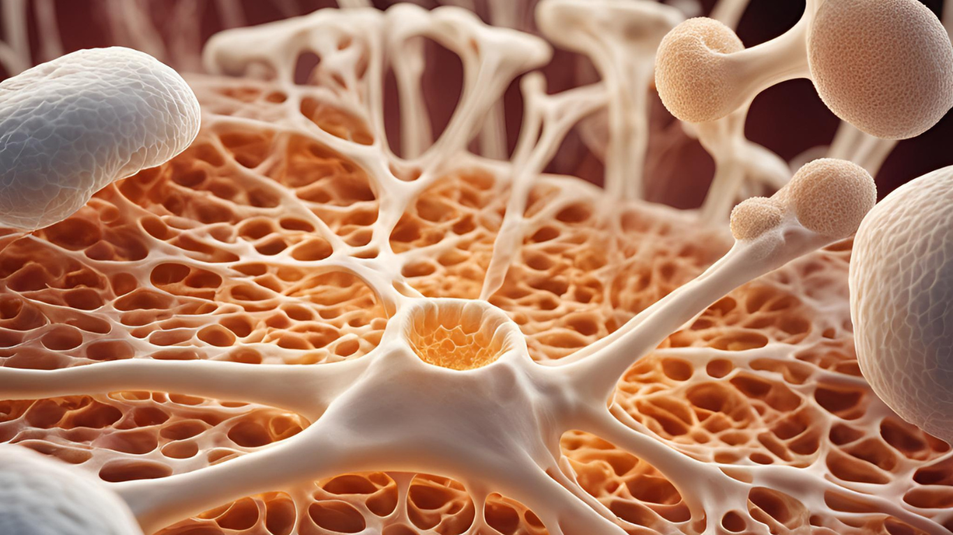
Ever wondered how bones actually form? Let’s explore the journey of bone formation, focusing on intramembranous and endochondral ossification. From early development in fetal life to the continuous process in adulthood, bone formation is a detailed and impressive process.
Ossification, or bone formation, starts in fetal life when existing connective tissue is replaced. Intramembranous ossification refers to the direct formation of bone from primitive mesenchyme. This process takes place in the clavicle and the bones of the skull vault. These membrane bones are formed through intramembranous ossification, providing the framework for the skeletal system. When ossification replaces a cartilaginous model, it is known as endochondral ossification. Bone formation begins at specific locations where the bone will develop, starting at the primary center of ossification.
- growth hormone,
- thyroid hormone,
- sex hormones.
Intramembranous ossification
In the region where the bone will form, mesenchyme becomes highly vascularized, and the cells begin to divide rapidly. Some cells in the center grow larger, become more basophilic, and differentiate into osteoblasts. These osteoblasts secrete a matrix known as the osteoid matrix. As more matrix is produced, the cells and their extensions become trapped within it. At this stage, the cells are called osteocytes.
The initially formed matrix contains collagen fibres and ground substance, giving it a soft structure. This osteoid matrix, which is unmineralized, quickly undergoes calcification to form bone. Cell division ensures a continuous supply of osteoblasts on the matrix surface. Meanwhile, nearby mesenchyme also transforms into osteoblasts to support ongoing bone formation.
Endochondral ossification
The hyaline cartilaginous model forms during embryonic life. The process starts in the center of the shaft in the diaphysis. It is called the primary center of ossification. This process leads to the formation of thin fenestrated plates, which become calcified. The calcified matrix reduces the diffusion of nutrients through it. As the cells do not get nutrition, chondrocytes degenerate and die leaving large interconnecting spaces.
At the same time
Perichondrium becomes an osteogenic layer. Osteogenic cells create a layer of osteoid matrix around the calcified cartilage. This layer is known as the periosteal collar. The bone thus formed becomes thickened and lengthens as ossification proceeds, and maintains the strength of the shaft. The perichondrium now becomes the periosteum. Vascular periosteal tissue, known as periosteal buds, start invading the calcified cartilage. These buds contain blood vessels and osteogenic cells, which can transform into osteoblasts. Meanwhile, osteoclasts erode the calcified cartilage after the cartilage cells die. As a result, thin partitions or trabeculae between lacunae break down, forming cavities. These cavities then become primary marrow spaces.
Afterwards, the osteoblasts arrange themselves on the surface of the calcified cartilage remnants. They lay down osteoid matrix on the remnants of the cartilage partitions, and this matrix eventually becomes mineralized. As a result, the earliest trabeculae have a core of cartilage covered by a layer of bone. Osteoclasts remove calcified cartilage. As a result, the cavity expands. The medullary or marrow cavity develops in the shaft. Support of the hone is provided by continued development of the periosteal collar.
From the primary ossific centre, the process of bone formation extends towards the end of the model. The cartilage that remains at the end of the bone is the epiphysis. This continues to grow by interstitial growth. This growth results in an increase in length of the model. Secondary ossification centres or the epiphyseal centres usually form after birth in the epiphyses. Bone formation extends from these centres in all directions. At the extreme ends, a layer of cartilage remains as the articular cartilage.
Epiphyseal plate
A plate of cartilage persists between the bone formed by the epiphyseal centre and that formed by the diaphyseal centre. This growth plate ensures the bone grows in length. It does this by proliferating cartilage at the epiphyseal end. As the cartilage degenerates, bone is deposited at the diaphyseal end. Thus, several zones of activity can be recognized in the epiphyseal plate.
Zones of Ossification

From the Epiphyseal end, the following zones are present.
Zone of reserve cartilage
This area is made up of hyaline cartilage. It is initially long, but shortens as the process of ossification encroaches it. There is slow growth in the region.
Zone of proliferation
In this zone, there is active proliferation of chondrocytes. These cells, in flattened lacunae, arrange in columns, separated by a small amount of matrix. Consequently, the continued interstitial growth of the cartilage in this zone is the mechanism by which bone increases in length.
Zone of maturation
In this zone there is cessation of cell division. Cells increase in size.
Zone of hypertrophy and maturation
Cells enlarge and accumulate glycogen in the cytoplasm. Lacunae are also becoming enlarged. The matrix separating cells and lacunae reduces to thin longitudinal partitions and horizontal septa. As the matrix calcifies, the cells die, and spaces form where the cells once were.
Zone of ossification
Vascular mesenchyme containing osteogenic cells invades the calcified matrix. Osteoblasts lay down bone on the surface of the calcified cartilage. Osteoclasts erode the calcified cartilage, and the marrow cavity increases in size. The part of the diaphysis next to the epiphyseal plate, where the body lays down bone, is the metaphysis. Growth in length stops when the bone completely replaces the epiphyseal plate. The zone of union forms the epiphyseal line.
Formation of osteons
The body initially lays down bone in the form of irregular plates. Collagen fibers arrange randomly in a network around scattered vascular spaces, without any organized layers. This type of bone, called woven fiber bone, occurs in developing fetal bones. It also appears in bones formed during fracture repair and around tooth sockets. Over time, the body begins to deposit roughly concentric layers of non-lamellated, parallel fiber bone. These layers form irregular Haversian systems, also called primary osteons, along the walls of narrowed vascular spaces. As the bone matures, typical Haversian systems or secondary osteons develop.
The formation of secondary osteons starts with the erosion of the earlier woven fiber bone and primary osteons. The body then deposits concentric lamellae of parallel fiber bone along the walls of the resorption cavity. A basophilic cement line, made of ground substance, marks the boundary between the eroded area and the newly deposited bone. This cement line is clearly visible between secondary osteons and the older bone.
As bone growth continues, erosion can also affect secondary osteons, leading to the formation of new Haversian systems. Remnants of earlier osteons persist as interstitial lamellae. The body also deposits circumferential lamellae on the surfaces of the periosteum and endosteum. These lamellae consist of parallel fibers. In one lamella, fibers run lengthwise. In the adjacent lamella, they run circularly. This arrangement provides strength and stability to the bone structure.
Remodelling of bone
Internal remodelling occurs throughout life, with the removal of old osteons and deposition of new ones. Such a process allows the bone architecture to change in response to altered mechanical stresses. Gross remodelling with changes of shape is seen in the bones of the skull. This process involves an increase in thickness. It also increases the surface area and curvature. Growth at the margins would increase the surface area. Differential rates of periosteal bone formation externally and bone erosion internally would result in an increase in thickness. The shaft of a long bone increases in diameter by periosteal deposition along with the endosteal erosion.
Repair of fractures
When a bone fractures, a blood clot forms at the site. Capillary loops and mesenchymal cells invade the clot, and the body lays down collagen, forming granulation tissue. Mesenchymal cells differentiate into chondroblasts and osteoblasts. The fibrous granulation tissue is replaced by hyaline cartilage. Woven fiber bone forms a provisional callus, which the body strengthens by depositing calcium. Osteogenic cells of the endosteum and periosteum also create a mesh work of woven bone. This occurs within and around the provisional callus, forming a bony callus. Later, osteoclastic and osteoblastic activity lay down lamellar bone at the site of the fracture, restoring the original form.


Leave a Reply