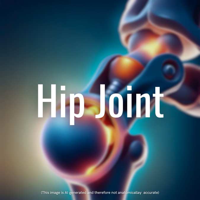
The hip joint is a synovial joint known for its stability and mobility. It is a multi-axial ball and socket joint, allowing movement in multiple directions. In this article, we will explore the anatomy, ligaments, movements, blood supply, and nerve supply of the hip joint, as well as some important clinical considerations.
Anatomy of the Hip Joint
The hip joint consists of the acetabulum, a horseshoe-shaped socket, and the head of the femur, forming a two-thirds sphere. The acetabulum is covered by hyaline cartilage and deepened by the acetabular labrum, a fibro cartilaginous rim. The transverse acetabular ligament bridges the acetabular notch and gives origin to the ligamentum teres.
Capsule and Synovial Membrane
The hip joint is surrounded by a capsule that attaches proximally to the margin of the acetabulum, including the transverse acetabular ligament, and distally to the anterior intertrochanteric line and the base of the trochanters. The posterior attachment is located 0.5 inches proximal to the intertrochanteric crest. Retinacular fibers, reflected from the distal attachment, provide a pathway for the blood supply of the femur.
The synovial membrane lines the capsule and covers the non-articular surfaces. It communicates with a bursa located deep to the tendon of iliopsoas, bulging out between the iliofemoral and ischiofemoral ligaments.
Ligaments of the Hip Joint
Several ligaments contribute to the stability of the hip joint:
- Iliofemoral Ligament: This is the strongest ligament and has an inverted “Y” shape. It arises from the anterior inferior iliac spine and inserts at each end of the trochanteric line. It resists hyperextension and usually remains intact in posterior dislocation.
- Pubofemoral Ligament: Located inferiorly, it arises from the iliopubic junction and blends with the capsule.
- Ischiofemoral Ligament: This posterior ligament arises from the ischium and inserts onto the greater trochanter. It is the weakest of the three ligaments.
- Round Ligament (Ligamentum Teres): Attached to the fovea capitis, this ligament is located within the joint and plays a minimal role in stability.
Movements of the Hip Joint
The hip joint exhibits high stability while allowing various movements:
- Flexion: The flexor muscles include the iliacus, psoas, rectus femoris, sartorius, and pectineus.
- Extension: The extensor muscles involve the gluteus maximus and the hamstrings.
- Adduction: Muscles responsible for adduction include adductor longus, adductor brevis, adductor magnus, gracilis, and pectineus.
- Abduction: The gluteus medius, gluteus minimus, tensor fascia lata, and sartorius are the main abductors.
- Medial and Lateral Rotation: The main medial rotator is the iliopsoas, while the gluteus maximus, gemelli, obturators, quadratus femoris, and piriformis contribute to lateral rotation.
![]() Clinical Point: Hip Dislocation
Clinical Point: Hip Dislocation
Usually, dislocated backwards—which is produced by force applied along the femoral shaft when the hip is flexed, leads to damage to sciatic nerve. Due to its close relationship. (Foot drop)
When hip is in adducted position – backward dislocation without acetabular fracture
When hip is in abducted position—backward dislocation with fracture of posterior
![]() Clinical Point: Hip Dislocation vs. Fractures
Clinical Point: Hip Dislocation vs. Fractures
The medial rotation of the femur by the iliopsoas muscle is useful in identifying hip dislocation. In hip dislocations, the femur is medially rotated, while in femoral neck fractures, the femur is laterally rotated.
![]() Clinical Point : Congenital Dislocation
Clinical Point : Congenital Dislocation
*Congenital dislocation of the hip is more common than any other joint. This occurs when the upper margin of the acetabulum is developmentally deficient
Blood Supply and Nerve Supply
The hip joint receives its blood supply through various sources:
- Ligamentum Teres: The obturator artery supplies the hip joint through the ligamentum teres.
- Retinacular Arteries: These arteries arise from the trochanteric anastomosis and are particularly important in adults.
- Nutrient Artery: The nutrient artery of the femur also contributes to the blood supply of the hip joint.
![]() Clinical Point : Blood Pressure Measurement
Clinical Point : Blood Pressure Measurement
Blood pressure in the lower limb can be recorded from the popliteal artery, which is a branch of the femoral artery.
The hip joint is innervated by several nerves that pass through the joint, including the femoral nerve, obturator nerve, sciatic nerve, nerve to quadratus femoris, and superior gluteal nerve.
![]() Clinical Point : Referred Pain in Hip Joint
Clinical Point : Referred Pain in Hip Joint
*pain in the hip joint is referred to knee


Leave a Reply