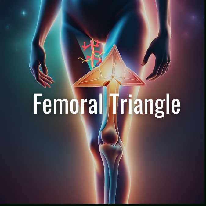
The femoral triangle is a triangular depression located on the front of the upper one-third of the thigh, just below the inguinal ligament. It serves as an important anatomical landmark in the thigh region. Let’s explore the boundaries, contents, and clinical significance of the femoral triangle.
Definition and Boundaries
- Definition: Triangular depression on the front of the upper one-third of the thigh, immediately below the inguinal ligament.
- Superior/Base: Inguinal ligament.
- Lateral: Medial border of the Sartorius muscle.
- Medial: Medial border of the Adductor Longus muscle.
- Apex: Point where the medial and lateral boundaries meet, directed downwards.
- Continuous below with the adductor canal.
| Boundaries | Description |
|---|---|
| Superior/Base | Inguinal ligament |
| Lateral | Medial border of Sartorius |
| Medial | Medial border of Adductor Longus |
| Apex | Point where the medial and lateral boundaries meet, directed downwards Adductor canal |
Floor (Medial to Lateral)
- Adductor Longus muscle
- Pectineus muscle
- Psoas major tendon
- Iliacus muscle
Roof
- Deep fascia (Fascia Lata): Contains the saphenous opening with cribriform fascia.
- Superficial fascia: Includes the upper part of the Great Saphenous vein, superficial inguinal nodes, and branches of the genitofemoral and ilioinguinal nerves.
- Skin
Contents
- Femoral Artery and Branches (from mid inguinal point to the apex):
- Superficial branches: Superficial external pudendal, superficial epigastric, superficial circumflex iliac.
- Deep branches: Profunda femoris, deep external pudendal, muscular branches.
(The femoral artery is commonly used for arterial blood gas sampling, catheterization, and checking femoral pulse.)
- Femoral Vein and Tributaries:
- Great saphenous vein (at the base, medial to the artery).
- Veins corresponding to branches of the artery (at the apex, posterior to the artery). It receives the greater saphenous vein, circumflex veins, and corresponding branches of the femoral artery.
- Femoral Sheath
- Nerves:
- Femoral nerve (located in the groove between the iliacus and psoas muscles, outside the femoral sheath).
- Nerve to the pectineus (branch of the femoral nerve).
- Femoral branch of the genitofemoral nerve.
- Lateral cutaneous nerve of the thigh: Compression can lead to meralgia paresthetica, resulting in numbness, tingling, and pain along the lateral thigh. Treatment may involve incising the inguinal ligament.
- Deep Inguinal Lymph Nodes
- Clinical significance: The femoral artery lies in front of the psoas major tendon, the femoral vein lies in front of the pectineus muscle, and the femoral nerve lies in the groove between the iliacus and psoas muscles.
Understanding the anatomical structures and clinical implications of the femoral triangle is essential for healthcare professionals in diagnosing and treating conditions related to the femoral region.


Leave a Reply