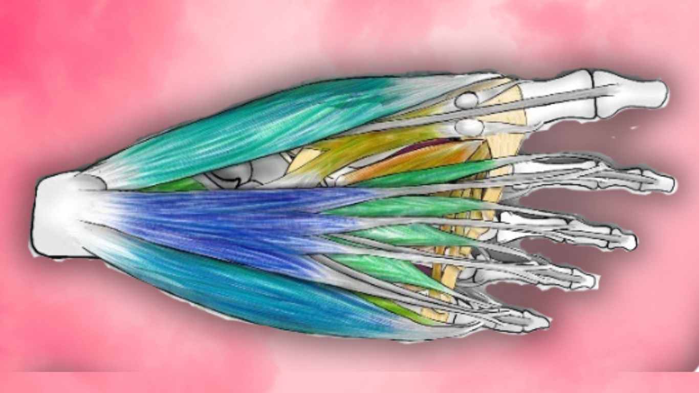
There’s a bundle of muscles piled, one on top of the other, in the sole of the foot. But, for convenience, it is described in four layers starting from the outermost and working your way in… just like you would in an actual dissection. There are 4 muscle layers of the foot, and this article expands on each of the muscles in these layers, in detail. For better understanding of the function of these muscles, read about the arches of the foot.
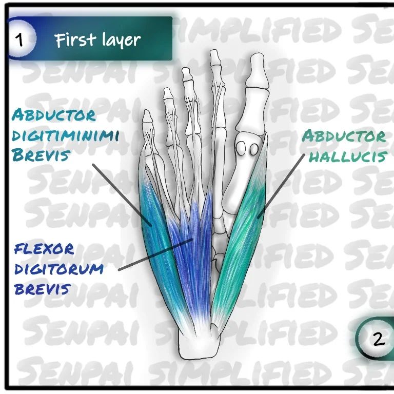
1st Layer
The first muscle layers of the foot consists of 3 muscles.
- Abductor hallucis
- Flexor digitorum brevis
- Abductor digiti minimi
The abductor hallucis originates from the medial tuberosity of calcaneum and flexor retinaculum. And it inserts on the base of the proximal phalanx of the big toe.
The flexor digitorum brevis also arises from the medial tubercle of calcaneum. This divides into four tendons and inserts into the border of the middle phalanx. In the four lateral toes. These tendons are perforated by the tendons of the flexor digitorum longus. The abductor digiti minimi, this is the one that flexes the pinky toe. It originates from both the medial and lateral tubercles of the calcaneum and inserts into the base of the proximal phalanx of the 5th toe.
The abductor hallucis can flex and abduct the big toe. And this supports the medial longitudinal arch. The flexor digitorum brevis, flexes the other four toes. This muscle supports both the medial and lateral longitudinal arches. The abductor digiti minimi can flex and abduct the 5th toe, and it supports the lateral longitudinal arch
Both abductor hallucis and flexor digitorum brevis are innervated by the medial plantar nerve. Abductor digiti minimi is innervated by lateral plantar nerve. When you dissect and remove these muscles, then you reach the second layer.
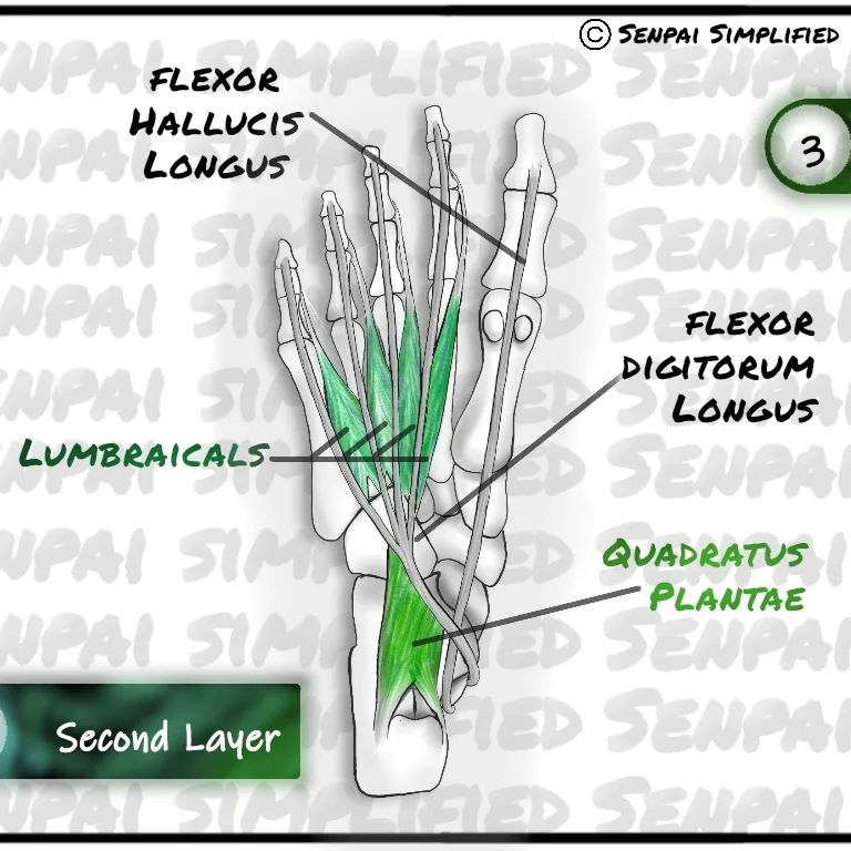
2nd Layer
The second layer of the muscle layers of the foot, has two muscles And two tendons of muscles that originate higher up, in the leg.
- The two muscles are,
- quadratus plantae (also known as flexor digitorum accessorius)
- 4 lumbraical muscles.
- The two tendons are,
- Flexor hallucis longus
- Flexor digitorum longus.
Quadratus plantae
The quadratus plantae originates from the medial and lateral sides of the calcaneum and inserts into the tendon of flexor digitorum longus. This muscle assists the flexor digitorum longus in flexing the lateral four toes. The flexor digitorum longus tendon lies kinda obliquely in the foot and the quadratus plantae sort of helps it to balance out the forces, otherwise it would be pulling the toes partly to one side. The lateral plantar nerve innervates this muscle.
4 Lumbraicals
The four lumbricals attach to the four tendons of the flexor digitorum longus. And distally, to the extensor expansion and the bases of the proximal phalanges of the lateral four toes. And this works exactly like the lumbraicals in the hand, it keeps the middle phalanx while bending the proximal phalanx down. The medial plantar nerve innervates the first lumbrical, while the lateral plantar nerve innervates the rest.
2 Tendons
The flexor longus tendon muscle that crosses underneath the flexor dogitorum longus tendon. An important thing to note here is that the lower limb has only one flexor digitorum muscle, but the upper limb has a flexor digitorum superficialis and a profundus. However, both the upper and lower limbs, there are two of each pollicis or the hallucis muscle. They are the longus and the brevis. Moreover, in both cases, the longus muscle attaches to the base of the distal phalanx, and the brevis muscle attaches to the base of the middle phalanx.
You can also note that the distribution of nerve supply of the medial and lateral plantar nerves is similar to the distribution of nerve supply by median nerve and ulnar nerve in the ventral side of the hand. When we remove this layer, we can reveal the 3rd layer.
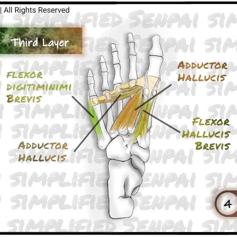
3rd Layer
This has 3 muscles.
- Flexor hallucis brevis
- Adductor hallucis, and
- Flexor digiti minimi brevis.
Abductor Hallucis
Let’s first talk about the abductor hallucis, This has 2 heads. The oblique head, and the transverse head. The oblique head arises from the bases of the second, third and fourth metatarsal bones. The transverse head, arises from the plantar ligaments. These two heads converge together and insert into the lateral side of the base of the proximal phalanx of the big toe.
Flexor Hallucis Brevis
The flexor hallucis brevis originates from the cuboid and lateral cuneiform, attaching to the tendon of the tibialis posterior at its insertion. This muscle divides into two heads towards the insertion. And divides into two tendons that insert into the medial and lateral sides of the base of the proximal phalanx of the big toe.
Flexor Digiti Minimi
And the last muscle of the 3rd layer, flexor digiti minimi. As the name explains it, this is the muscle that flexes the small toe. It arises from the base of the fifth metatarsal bone and inserts into the base of the proximal phalanx of the 5th toe, on its lateral side. The lateral plantar nerve innervates this muscle.
Innervation
The medial plantar nerve innervates the flexor hallucis brevis, while the deep branch of the lateral plantar nerve innervates the abductor hallucis.
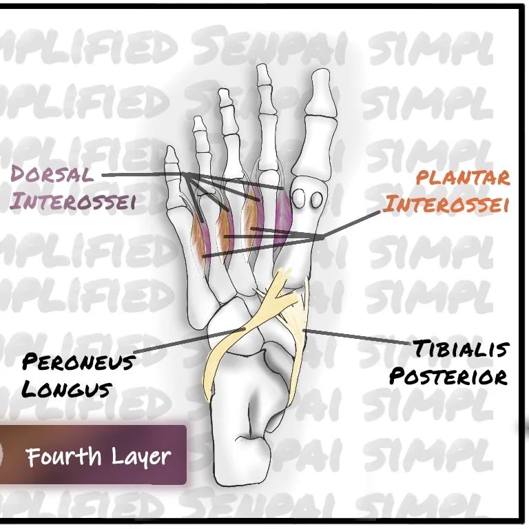
4th Layer
The 4th layer is similar to the second layer because this also has two tendons and two muscles.
- The two muscles are
- The plantar interossei
- And Dorsal interossei
- The two tendons are
- tibialis posterior
- peroneus longus.
There are 3 plantar interossei muscles. They originate from the 3rd fourth and 5th metatarsals, from their inferior surfaces. And they insert on the medial side of the base of the proximal phalanges of the 3rd, 4th and 5 toes respectively.
The tibialis posterior originates higher up in the leg and tendon reaches the foot and divides into two. The larger superficial division inserts onto the plantar surface of the tarsal bones of the foot, mainly onto the tuberosity of the navicular bone, and the medial cuneiform bone. The smaller deeper division inserts onto the middle and lateral cuneiform bones, the cuboid and second third and fourth metatarsals.
Summary of muscle layers of the foot
| Layer | Muscle | Origin | Insertion | Innervation |
|---|---|---|---|---|
| 1 | Abductor hallucis | Medial tuberosity of calcaneum, flexor retinaculum | Base of proximal phalanx of big toe | Medial plantar nerve |
| 1 | Flexor digitorum brevis | Medial tubercle of calcaneum | Border of middle phalanx of lateral four toes | Medial plantar nerve |
| 1 | Abductor digiti minimi | Medial and lateral tubercles of calcaneum | Base of proximal phalanx of 5th toe | Lateral plantar nerve |
| 2 | Quadratus plantae | Medial and lateral sides of calcaneum | Tendon of flexor digitorum longus | Lateral plantar nerve |
| 2 | Lumbricals | Four tendons of flexor digitorum longus | Extensor expansion and bases of proximal phalanges | Medial and lateral plantar nerves |
| 2 | Flexor hallucis longus | Higher up in the leg | Base of distal phalanx of big toe | Tibial nerve |
| 2 | Flexor digitorum longus | Higher up in the leg | Distal phalanges of lateral four toes | Tibial nerve |
| 3 | Flexor hallucis brevis | Cuboid, lateral cuneiform | Base of proximal phalanx of big toe, medial and lateral sides | Medial plantar nerve |
| 3 | Adductor hallucis | Bases of second, third, and fourth metatarsal bones, plantar ligaments | Lateral side of base of proximal phalanx of big toe | Medial plantar nerve |
| 3 | Flexor digiti minimi brevis | Base of fifth metatarsal bone | Base of proximal phalanx of 5th toe, lateral side | Lateral plantar nerve |
| 4 | Plantar interossei | Inferior surfaces of 3rd, 4th, and 5th metatarsals | Medial side of base of proximal phalanges of 3rd, 4th, and 5th toes | Lateral plantar nerve |
| 4 | Tibialis posterior | Higher up in the leg | Plantar surface of tarsal bones, tuberosity of navicular bone, medial cuneiform bone, middle and lateral cuneiform bones, cuboid, and second, third, and fourth metatarsals | Tibial nerve |
| 4 | Peroneus longus | Higher up in the leg | Base of first metatarsal, medial cuneiform, and base of second metatarsal | Superficial peroneal nerve |


Leave a Reply